Wednesday, February 22, 2006

Shin Splints and Anterior Leg Pain- A Synopsis

What exactly are “Shin Splints”?

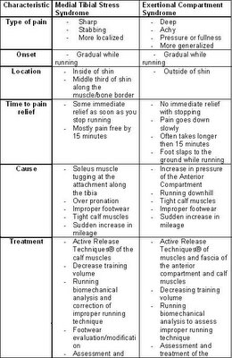
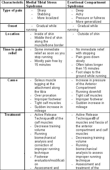
The term “shin splints” is a common diagnosis given when someone is suffering from pain in the front of their legs and is often associated with running. There are two main types of shin splints. They both have very similar symptoms but are significantly different in their actual diagnosis and treatment. So how can you tell them apart? Let's start with a description of both and then compare them…

FIRST TYPE
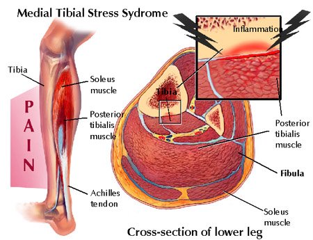
Medial Tibial Stress Syndrome (MTSS), as the name describes, is caused by pain along the medial (inside) part of the tibia (shin bone). The pain typically develops over a steady time while running and can be painful enough to make you want to stop. The pain is sharp and it decreases significantly once you stop running and after about 15 minutes is almost completely gone. You will also find that your shins are tender or painful to the touch along the middle third of the inside of the tibia. This type of shin splint usually begins with the onset of a new running activity and/or a sudden or rapid increase in mileage. An increase in body weight and running on hard surfaces has also been known to lead to this type of shin pain.
So what causes this pain? It is caused by the Soleus Muscle that attaches to the tibia along its inside border.


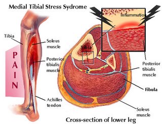
This muscle lies deep to the Gastrocnemius (bigger, bulkier muscle on the back of the leg) and together they form the “calf muscles”. The Soleus is composed of slow twitch muscle fibers, which means it is involved in endurance activities such as running, walking and even maintaining your standing posture. Once this muscle gets tight and/or overworked from sudden increases in running mileage or when starting a new activity, the muscle begins to tug at the attachment along the medial border of the tibia. This is what ends up causing the pain on the inside of the shin. The body naturally wants to try to stop the painful tugging, so it lays down scar tissue along the attachment to help reinforce it. However this only causes the muscle to become more tight and places even more stress along the attachment at the shin. This creates a viscious cycle of pain and tightening that will continue either until you get treatment, stop the activity, or modify the activity to provide enough time for proper healing.

SECOND TYPE
The second type of shin splints is also associated with a sudden increase in running mileage, and/or the beginning of a new running activity. The pain in with this type of shin splint is often worse running down hill, the pain is a deeper, achy pain and can lead to a slapping foot while running. Once you stop running the pain does not go away immediately and can still be very painful at rest 15 minutes after stopping. This time the pain is lateral (on the outside) of the tibia and is often described as a fullness or pressure feeling in the leg and is called an Exertional Compartment Syndrome.

The cause of the pain in this scenario is an increase in pressure in the anterior compartment of the leg. This compartment is formed between the tibia and fibula (the two bones in the lower leg) and a thick layer of fascia around the front. In this compartment lies the Tibialis Anterior muscle as well as the muscles that extend your toes. When you are running, these muscles help to lift (dorsiflex) your foot and toes to allow ground clearance while in the swing phase of running. They also help to slowly lower your foot and toes to the ground after heel strike at the beginning of the stance phase of running. When you contract a muscle, there is an increased need for blood to supply enough nutrients to keep it working. This increased blood supply to the muscle in turn increases the size of the muscle. This process is normal and usually goes unnoticed, however if the size or volume of the muscle increases too much, especially when the muscle is held tight like in the anterior compartment, it results in an increase in pressure and this causes pain. The pressure in the anterior compartment can get high enough that it affects the muscles ability to work, and this is why you may experience your foot slapping the ground while running. This results because the muscles can no longer lower your foot and toes slowly to the ground once you have heel contact so they just slap down uncontrollably. If the pressure continues to go up, it can even shut off the sensory nerve contribution to the skin between your first two toes.


Treatment Options
In order to decrease the pain associated with MTSS, the amount of pulling that the Soleus muscle exerts on the tibia must be decreased. This can be accomplished in several ways. The first is by treating the Soleus with muscle soft tissue techniques such as ART® and Graston. These techniques have been developed to help break up scar tissue, release adhesions between muscles and restore normal muscle tone. By using these techniques, the tension on the tibial attachment of the Soleus is lowered thereby decreasing the pain at that area. More treatment options include walking/running biomechanical analysis. Performed properly, this can reveal functional mechanics that may be predisposing to MTSS. Some predisposing factors include overpronation, rearfoot varus deformity and leg length discrepancy. If these areas are addressed, the time to recovery and recurrence rates can be greatly decreased. Footwear and training program analysis should also be performed to make sure they are tailored to your specific needs. It is recommended that you do not increase mileage by more then 10% per week in order to decrease the likelihood of developing MTSS.

The treatment options for ECS are similar to those for MTSS since poor running mechanics, improper footwear, and tight muscles can predispose you to being susceptible to this type of injury. More specifically, performing ART®, Graston, Medical Acupuncture, and other soft tissue techniques over the Tibialis Anterior, Calf Muscles, foot and toe extensors as well as the compartment fascia can help lower the pressure in the compartment and therefore decrease the pain associated with it.

Considerations
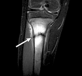
Two serious conditions can also be associated with common “Shin Splints”. The first results if a MTSS goes untreated for a long time and the activity is not modified appropriately. Then there is a chance of developing a tibial stress reaction. This Stress Reaction can lead to a visible stress fracture on X-ray which, if present, requires about 6-8 weeks of restricted physical activity and significant rehabilitation.


A more severe condition that may develop is called an Acute Anterior Compartment syndrome. This is a medical emergency since the pressure in the compartment can go so high that it cuts off blood and nerve supply to the muscles, ligaments, and bones in the lower leg and foot. The treatment is a surgical procedure called a fasciotomy. A fasciotomy involves cutting open a 4-6 inch section of the anterior compartment to decrease the pressure. Symptoms of this condition are extreme pain, swelling, numbness and lack of muscle control in the foot. These symptoms are similar to those of ECS except they are more intense and DO NOT decrease in intensity - they only INCREASE with time!


Summary


If you currently experience any of these symptoms, you should see a qualified health care practitioner to examine and evaluate your specific condition.


Disclaimer
This information is not intended to be a substitute for professional medical advice. You should not use this information to diagnose or treat a health problem or disease without consulting with a qualified health care provider. Please consult your health care provider with any questions or concerns you may have regarding your condition. Any attempt to diagnose and treat an illness using the information in this site should come under the direction of a trained medical practitioner. We accept no responsibility for any adverse effects or consequences resulting from the use of any of the suggestions or procedures in this site or related internet links. By using the information in this web site you are confirming that you understand this statement and that you accept all risk and responsibility.
All matters regarding your health should be supervised by your health care provider. All information provided in this site is for the purpose of education, not diagnosis or treatment.
Rotator Cuff Injuries and the S.I.C.K. Scapula

Do you suffer from a chronic rotator cuff injury?

The problem may not be the rotator cuff, you might have a S.I.C.K. scapula!

Here's what to look for:

Are you an Overhead Athlete? (Baseball, Volleyball, Tennis…)
Do you have heaviness in your arm with throwing/overhead activities?
Have you ever experienced “Dead Arm”?
Do you suffer from “Shoulder Impingement” or a Rotator Cuff injury?
Have you ever been diagnosed with a SLAP lesion?

If you answered yes to any of these questions, you may have a S.I.C.K. Scapula!
Often diagnosed as a rotator cuff injury, the following will help to illustrate why you may not be getting better with simple rotator cuff treatment and rehabilitation.

The S.I.C.K. scapula is a pneumonic that was developed to help describe a group of findings in the shoulder and scapula (shoulder blade). S.I.C.K. scapula stands for Scapular malposition, Inferior medial scapular winging, Coracoid tenderness, and scapular dysKinesis. First put forward by a group of authors to describe conditions observed in professional baseball players, this new approach to addressing shoulder problems is changing the way shoulder treatment and rehabilitation is viewed. This will all be described in the following article.

First thing to know…Anatomy of the shoulder!


The scapula, commonly called your shoulder blade is the only attachment of your arm to the rest of your body. The scapula is also the origin of very important muscles that move the arm, such as the rotator cuff (Supraspinatous, Infraspinatus, Teres Minor, and Subscapularis muscles) and 9 other muscles that do a variety of work to help you shrug your shoulders, move your arms and even your forearms. The scapula has only one attachment to another bone and that is your clavicle (or collarbone). The clavicle provides the only solid bony attachment of your hand, forearm, and shoulder to the rest of your body!

How can one little bone like your clavicle be responsible for allowing and supporting so much movement in the shoulder, arm, and hand?

The answer is that it has support from a number of muscles that help hold and stabilize your shoulder blade on the back of your rib cage. This becomes very important since without a solid attachment on your body, the shoulder doesn't have a strong base of support to work from and you can get pain/injuries while performing simple tasks like throwing a ball, working overhead or even just doing a push-up.

So how does all this cause the pain you get in your shoulder? According to the work done by Burkhart, Kibler, et al., what they found in professional baseball players with shoulder pain was a greatly decreased range of motion in internal rotation of the throwing shoulder. They linked the decrease in internal rotation with an increase in external rotation and tightening of part of the shoulder capsule. The tightening of the posterior shoulder capsule actually changes the axis of rotation of the shoulder, allowing for the increase in external rotation and decrease in internal rotation. This change in the axis of rotation and resulting increase in external rotation provides an opportunity for the development of a SLAP lesion (Superior Labrum Anterior Posterior). A SLAP lesion is damage to the cartilage (labrum) that helps deepen your shoulder joint. This occurs because the long head of the Biceps Muscle in your arm attaches in this cartilage (labrum) and with more external rotation available in the shoulder, the tendon of the biceps can actually peel this cartilage off of the bone resulting in a SLAP lesion and a significant amount of pain and discomfort.

Another finding in these professional athletes was that they had what is called scapular winging. Scapular winging is when the bottom tip and/or the inside border of the shoulder blade sticks out, or wings away from the rib cage at rest and is exaggerated with certain arm movements. This is often caused by having weak muscles that hold the scapula on the rib cage, most commonly the middle and lower trapezius and the serratus anterior. In addition to weak muscles, there are often tight muscles that help “pull” the scapula off the rib cage. The main muscle that does this is the Pectoralis Minor. This muscle attaches from the ribs on your chest to the coracoid process of the scapula (which is on the top and front of the shoulder just below and deep to the collarbone). When this muscle gets tight, it pulls the scapula up and over the top of the rib cage, resulting in this inferior-medial border winging.

In addition to causing scapular winging, this change in position also causes another change in the axis of rotation of the shoulder joint resulting in early pinching of the humerus against the top of the scapula. This early pinching is most noticeable by raising your arms straight up in front of you. If you get pain in the shoulder, it is sometimes called an impingement syndrome. Now if you push your shoulder blade firmly against your back by leaning on a wall and try raising your arm in front of you, you should get it higher before you feel pain. This is because by pressing your shoulder blade onto your rib cage, you are restoring the proper position of the shoulder blade resulting in a more normal axis of rotation and thereby restoring normal range of motion.

A final finding that was noticed in this group of athletes was that the scapula, which normally moves in a smooth fashion when you raise your arms is now not moving properly. This is called Scapular Dyskinesis. With normal shoulder movement, the inferior tip of the scapula moves outward and upwards. In the SICK scapula, the inferior medial tip does not move in the same fashion as the healthy shoulder and gets “stuck” in a lower position.

As you can see, if you have been doing weeks and months of rotator cuff strengthening exercises with rubber tubing and you still have shoulder pain, you may need to address the muscles that help hold that shoulder blade on your ribcage. Without a strong base of support to work from, the rotator cuff will not be able to fully recover. The shoulder and arm can only get as strong as the muscles that hold the shoulder blade onto your body.

Lets now summarize our S.I.C.K. scapula pneumonic:
S: Scapular malposition on the rib cage
I: Inferior medial border winging from weak middle and lower trapezius muscles
C: Coracoid pain and malposition from the attachment of a tight Pectoralis minor muscle pulling on the coracoid
K: Scapular dyskinesis from alterations in muscle recruitment patterns

Many of the above findings are also present in recreational athletes, office and computer workers. The typical posture of a computer operator is with the head tilted down, shoulders rolled forwards and arms stretched out in front of them. This allows for tightening of the pectoralis minor on the front of the body since the shoulders are already rolled forward. It also allows for the lower trapezius muscle to get weak because of lack of use from not holding your shoulders back. As a result of these tight and weak muscles, the shoulder blades don’t sit properly on the rib cage and when you move your arms, you have alterations in the proper movement patterns. Now you can see how not only professional athletes can suffer from this type of condition. As a result, many people suffer from shoulder pain that is a result of poor shoulder position on the rib cage and from altered muscle firing patterns.

How should this be treated?

The best treatment for the elite professional athlete is physical rehabilitation. The authors found that by stretching the posterior shoulder capsule, strengthening the muscles that hold your scapula in place and stretching/relaxing the tight muscles that pull the scapula into a different position.

Many treatment forms are available for treating this type of condition. Soft tissue techniques such as Active Release Techniques (ART®), Graston, and Medical Acupuncture are designed to address these issues on functional, anatomical, and neurological levels.

Be sure to have a complete physical examination by a qualified healthcare practitioner in order to receive a proper diagnosis and plan of management.


Here is a sample of a few of the rehabilitation exercises that should be performed by someone with a S.I.C.K. scapula as outlined by Burkhart, Morgan and Kibler:


2. Scapular Clock Exercise


References:

Burkhart SS, Morgan CD, Kibler WB. The disabled throwing shoulder: spectrum of pathology Part I: pathoanatomy and biomechanics. Arthroscopy. 2003 Apr;19(4):404-20

Burkhart SS, Morgan CD, Kibler WB. The disabled throwing shoulder: spectrum of pathology. Part II: evaluation and treatment of SLAP lesions in throwers.Arthroscopy. 2003 May-Jun;19(5):531-9

Burkhart SS, Morgan CD, Kibler WB. The disabled throwing shoulder: spectrum of pathology Part III: The SICK scapula, scapular dyskinesis, the kinetic chain, and rehabilitation.Arthroscopy. 2003 Jul-Aug;19(6):641-61

Kibler WB, Uhl TL, Maddux JW, Brooks PV, Zeller B, McMullen J. Qualitative clinical evaluation of scapular dysfunction: a reliability study. J Shoulder Elbow Surg. 2002 Nov-Dec;11(6):550-6.
Rubin BD, Kibler WB. Fundamental principles of shoulder rehabilitation: conservative to postoperative management. Arthroscopy. 2002 Nov-Dec;18(9 Suppl 2):29-39.

Kibler WB, McMullen J. Shoulder rehabilitation strategies, guidelines, and practice. Orthop Clin North Am. 2001 Jul;32(3):527-38

Kibler WB, Livingston B. Closed-chain rehabilitation for upper and lower extremities. J Am Acad Orthop Surg. 2001 Nov-Dec;9(6):412-21

Kibler WB, McMullen J. Scapular dyskinesis and its relation to shoulder pain. J Am Acad Orthop Surg. 2003 Mar-Apr;11(2):142-51


Disclaimer
This information is not intended to be a substitute for professional medical advice. You should not use this information to diagnose or treat a health problem or disease without consulting with a qualified health care provider. Please consult your health care provider with any questions or concerns you may have regarding your condition. Any attempt to diagnose and treat an illness using the information in this site should come under the direction of a trained medical practitioner. We accept no responsibility for any adverse effects or consequences resulting from the use of any of the suggestions or procedures in this site or related internet links. By using the information in this web site you are confirming that you understand this statement and that you accept all risk and responsibility.
All matters regarding your health should be supervised by your health care provider. All information provided in this site is for the purpose of education, not treatment.
Adolescent Idiopathic Scoliosis

What is it?
Scoliosis is a lateral curvature of the spine (> 10°) in the frontal plane with accompanying rotation of the vertebrae about a longitudinal axis. More specifically, adolescent idiopathic scoliosis (AIS) is a scoliosis of unknown origin that affects children between the ages of approximately 10 and 18 years of age.

Who gets it?
AIS affects approximately 2 percent of children between the ages of 10 and 16. AIS of small curvatures occurs with equal rates in both boys and girls in that age group. However, curves of larger magnitudes (> 30°) occur ten times more often in females then in males. Females also have a greater risk of progression. The prevalence of curves greater than 30° is 0.2 percent, and curves greater than 40° is 0.1 percent.

What do we know about it?
The precise cause of AIS is still largely uncertain. Many theories exist which list genetics, skeletal growth, melatonin deficiency, neuromuscular abnormalities, and metabolic disturbances as possible causes.
- Thoracic curves with the apex on the right are the most common type of curve, followed by a double thoracolumbar curve which is the second most common.
- Only 10% of adolescents with scoliosis have curve progression requiring medical intervention.
- There are three main determinants of scoliotic curve progression: patient gender, curve magnitude at time of diagnosis, and future growth potential.
- Curves rarely progress once skeletal maturity is reached. Skeletal maturity can be determined using Risser’s sign.

Risser’s Sign:


Lonstien & Carlson:


Progression Factor =


How do we manage it?
Curves between 10-15 degrees require no active treatment, but should be monitored every 6 months.
- Curves between 25-40 degrees should be considered for bracing.
- Curves over 30 degrees should be monitored by X-ray every 3 months.
- Curves over 40 degrees should be referred for surgery.
- Alternative to bracing is Lateral Electric Surface Stimulation. This form of treatment has been shown to produce similar results as bracing when measuring curve progression.

Treatment and Referral Guidelines for Patients with Scoliosis:


Other factors concerning scoliosis:
- The majority of curves spontaneously stabilize themselves and thereby reduce the risk of further curve progression.
- Controversy exists on whether patients with scoliosis have higher incidence of back pain than those without.
- There exists an association between AIS and decreased bone mineral density (osteopenia), however the underlying cause is not known and the curve magnitude does not appear to be linked with the degree of osteopenia.


References:

Cheng, J., Tang S.P., Guo, X. Chan, C., Qin, L. Osteopenia in Adolescent Idiopathic Scoliosis. Spine 2001, E19-E23.

Greiner, K.A. Adolescent Idiopathic Scoliosis: Radiologic Decision-Making. American Family Physician, Vol 65, No. 9, May 15, 2002.

Reamy, B., Joseph Slakey. Adolescent Idiopathic Scoliosis: Review and Current Concepts. American Family Physician, Vol 64, No. 1, July 1 2001.


Disclaimer
This information is not intended to be a substitute for professional medical advice. You should not use this information to diagnose or treat a health problem or disease without consulting with a qualified health care provider. Please consult your health care provider with any questions or concerns you may have regarding your condition. Any attempt to diagnose and treat an illness using the information in this site should come under the direction of a trained medical practitioner. We accept no responsibility for any adverse effects or consequences resulting from the use of any of the suggestions or procedures in this site or related internet links. By using the information in this web site you are confirming that you understand this statement and that you accept all risk and responsibility.
All matters regarding your health should be supervised by your health care provider. All information provided in this site is for the purpose of education, not treatment.
Tendinosis: History, Histopathology, Pathogenesis, Evaluation, and Treatments of the underreported chronic tendon condition.

The term tendinosis was first used in the 1940’s by a group of German researchers, however the term did not receive much attention until it was used again in the mid 1980’s to describe a non-inflammatory tendon condition. The more commonly used term of ‘tendinitis’ has since been proven to be a misnomer for several reasons. The first of which is that there is a lack of inflammatory cells in conditions that were typically called a tendonitis. Since inflammation is the key pathological process involved with that term, and the discovery that there in fact were no, or very few, inflammatory cells present in the condition, a new term was adopted – tendinosis. This has a much greater impact then just the name one calls a condition, it also has a profound impact on how the condition is treated. Khan compares the time to recovery of an acute and chronic presentation of tendinosis versus tendonitis, as well the prevalence and focus of conservative therapy. They are outlined as follows:


Histopathology of Tendinosis

A typical healthy tendon is composed of primarily type I collagen with minimal amounts of type III collagen interspersed within the neatly arranged parallel fiber orientation of type I collagen. The healthy tendon is said to be white and shiny and reflect polarized light under microscope. Unhealthy tendons, or tendinosis, appear gray and do not reflect polarized light under the microscope. As previously mentioned, there are no inflammatory cells found in the collagen of chronic tendinosis conditions, there may however be a few chronic (not acute) inflammatory cells present if there is healing of a partial tear of the tendon. There are however three key findings present in tendinosis conditions. They are: disrupted collagen fibers within the tendon, increased cellularity, and neovascularization.

Kraushaar and Nirschl found that on cross section of collagen, an area of tendinosis showed that the collagen was of variable diameter, uneven mixture of thick and thin fibrils and in some areas did not even connect with each other to form a tendinous structure. They concluded that the ultrastructure of collagen in tendinosis is unable to sustain a tensile load. Maffulli et al. confirmed these findings and also discovered that the collagen that is formed in an area of tendinosis is actually type III collagen, instead of the predominant type I in healthy tendon. This increase in type III collagen, and possible decrease in type I collagen results in a decrease in the forces that the tendon can withstand and may eventually lead to tendon rupture.

The other two findings present in tendinosis, increased cellularity and neovascularization has been termed angiofribroblastic hyperplasia by Nirschl. The cells present in tendinosis are mesenchyme-derived tendon fibroblasts (tenocytes) and myofibroblasts, as opposed to the humoral process of an immune based inflammatory response. These are cells that represent a degenerative condition. Khan described the types of degeneration associated with tendinosis as either mucoid or lipoid. Mucoid degeneration causes softening of the area affected as well as the characteristic changes seen in tendon from the glistening white appearance to the gray appearance in tendinosis. Lipoid degeneration is characterized by an abnormal increase in lipid material in a tendon.

Neovascularization found in tendinosis has been described as a haphazard arrangement of new blood vessels and Kraushaar et al. even mention that the vascular structures do not function as blood vessels. Vessels have even been found to form perpendicular to the orientation of the collagen fibers. They then concluded that the increased vascularity present in tendinosis is not associated with increased healing.


Pathogenesis

Tendinosis is said to be a degenerative condition affecting a tendon. The cause of this degeneration has been thought to be due to one of several things, of which overuse causing microtrauma appears to be the most widely accepted. Kraushaaar describes four stges of injury due to micro trauma. Stage-1 is said to be most likely inflammatory in nature and is associated with pathological alterations that most likely will resolve. Stage-2 injury is associated with pathological alterations such as tendinosis and angiofibroblastic degeneration. Stage-3 injury is associated with pathological changes (tendinosis) and complete structural failure (rupture). Finally, stage-4 injury exhibits features of both stages 2 and 3 along with other findings such as fibrosis, soft matrix calcification, and hard osseous calcification.

There is also said to exist a tendinosis cycle that must be broken before successful treatment and pain-free movements can be restored. The cycle of tendinosis is simplified and described as repetitive overuse activities or microtraumas causing microtears in the tendon creating a fibroblastic hyperplasia response, which then respond by increasing the amount of type III collagen within a tendon. This further weakens the tendon causing more microtears and prolonging the degenerative cycle.

Treatment for this condition must first attempt to stop the degenerative cycle and then to restore proper collagen synthesis, strength and function.


Conservative Management

Kraushaar et al. lists the primary goals of treatment of tendinosis as being pain control, preservation of motion, flexibility and strength, and the development of endurance over time. Since the exact cause of pain in tendinosis is not known, many theories have tried to explain the mechanism of pain in order to improve treatment. The original theory of pain is based back to the concept of tendonitis where inflammation is the source of pain. Since there are no inflammatory cells present in tendinosis, this theory no longer applies. The next theory to evolve was that it is the biomechanical changes that result in pain production. Since a disruption of collagen is painful in a ligament sprain, why can’t the disruption of the collagen fibers in a tendinosis also be the cause of pain? Khan argues this notion by comparing a few post surgical patients with patellar tendinopathy who experience pain against those who do not. He mentions that the area when visualized is indistinguishable between painful and pain-free subjects, thereby negating a relationship between pain and collagen status. The next theory involves a biochemical model of pain. It is hypothesized that certain unknown chemicals may be responsible for pain production within an area of tendinosis. Certain chemicals that are already proven to be pain stimulators are glutamate and chondroitin sulfate.

Traditional treatments involve the use of corticosteroid injections for the treatment of tendonitis. It has been proven that corticosteroid injections actually further the degeneration of tendons and increase the risk of recurrence of the condition as well as increase the risk of tendon rupture. That being said, corticosteroid injections have been shown to be effective in short term pain reduction for tendinosis. The mechanism is unclear but it is thought that bathing the area may alter or interfere with the local chemicals that cause the pain stimulus in the area.

Other treatments include biomechanical reduction of stress on the tendon, relative rest, and ice since it is a vasoconstrictor (and increased vascularity is a finding in tendinosis) and a natural analgesic. Research for the use of modalities such as laser and therapeutic ultrasound is controversial at best, however it is recommended by certain sources to help stimulate collagen synthesis. Stretching and strengthening are two common methods of treating tendinosis and eccentric strengthening is gaining popularity quite rapidly.

Khan states that eccentric strengthening results in a stimulation of mechanoreceptors in tenocytes to produce collagen and thereby help reverse the tendinosis cycle. Alfredson et al. used heavy load eccentric calf muscle training in the treatment of chronic Achilles tendinosis and the found that all 15 participants were back to their preinjury activities after the 12 week training period. They performed 3 sets of 15 repetitions, with both the knee straight and knee bent, twice daily for twelve weeks. They did not perform the concentric portion of the exercise with the injured leg, they relied on the healthy leg to return them to the starting position each time. A more recent study by Shalabi et al. found that eccentric strengthening of the gastrocnemius-soleus complex resulted in a decreased tendon volume and intratendinous signal when evaluated by MR imaging. They also concluded that the findings were associated with decreased pain scores on a VAS and is therefore an effective adjunct in the treatment of Achilles tendinopathies.

Take Home Points:

1.
Chronic tendon injuries are degenerative in nature and NOT inflammatory.
2. Anti-inflammatory medications (NSAID’s) and/or corticosteroid injections can actually accelerate the degenerative process and make the tendon more susceptible to further injury, longer recovery time and may increase likelihood of rupture.
3. Heavy load eccentric strength training helps to increase the tensile strength of the tendon, increase the amount of Type I collagen and decrease the amount of ground substance thereby reducing tendon volume and helping restore the tendon to a healthy state.


References:

Alfredson, H., Pietila, T., Jonsson, P., Lorentzon, R. Heavy-Load Eccentric Calf Muscle Training For the Treatment of Chronic Achilles Tendinosis. AJSM. Vol. 26 (3), 1998. pp.360-366

Cook, JL., Khan, KM., Maffulli, N., Purdam, C. Overuse Tendinosis, Not Tendinitis: Part 2: Applying the new approach to patellar tendinopathy. The Physician and Sportsmedicine, Vol 28 (6), June 2000.

Khan, KM., Cook, JK. Overuse Tendon Injuries: Where does the pain come from? Clinical Sports Medicine

Khan, KM., Cook, JL., Kannus, P., Maffulli, N., Bonar, SF. Time to abandon the “Tendinitis” Myth: Pianful, overuse tendon conditions have a non-inflammatory pathology. BMJ. Vol. 324 16 March 2002. pp.626-627.

Khan, KM., Cook, JL., Taunton, JE., Bonar, F. Overuse Tendinosis, Not Tendinitis: Part 1: A new paradigm for a difficult clinical problem. The Physician and Sportsmedicine, Vol 28 (5), May 2000.

Khan, KM. Cool, JL., Bonar, F., Harcourt, P., Astrom, M. Histopathology of Common Tendinopathies: Update and implications for clinical management. Sports Medicine. Jun 27 (6), 1999, pp. 393-408.

Kraushaar, B., Nirschl, RP. Tendinosis of the Elbow (Tennis Elbow). The Journal of Bone and Joint Surgery. Vol 81-A (2), Feb. 1999.

Maffulli, N., Ewen, SWB., Waterston, SW., Reaper, J., Barrass, V. Tenocytes from Ruptured and Tendinopathic Achilles Tendons Produce Greater Quantities of Type III Collagen than Tenocytes from Normal Achilles Tendons: An in vivo model of human tendon healing. AJSM Vol 28 (4), 2000.

Shalabi, A., Kristofferson-Wilberg, M., Svensson, L., Aspelin, P., Movin, T. Eccentric Training of the Gastrocnemius-Soleus Complex in Chronic Achilles Tendiopathy Results in Decreased Tendon Volume and Intratendinous Signal as Evaluated by MRI. AJSM, Vol. 32 No. X, 2004


Disclaimer
This information is not intended to be a substitute for professional medical advice. You should not use this information to diagnose or treat a health problem or disease without consulting with a qualified health care provider. Please consult your health care provider with any questions or concerns you may have regarding your condition.Any attempt to diagnose and treat an illness using the information in this site should come under the direction of a trained medical practitioner. We accept no responsibility for any adverse effects or consequences resulting from the use of any of the suggestions or procedures in this site or related internet links. By using the information in this web site you are confirming that you understand this statement and that you accept all risk and responsibility.
All matters regarding your health should be supervised by your health care provider. All information provided in this site is for the purpose of education, not treatment.